Smart responsive hydrogel-based growth factor delivery systems: precise release and synergistic regeneration strategies for wound healing
Authors
Kan Zhan, Yawen Xue, Junping Zhou, Yanan Xue, Liqun Jin, Renchao Zheng, Yuguo Zheng
- aKey Laboratory of Bioorganic Synthesis of Zhejiang Province, College of Biotechnology and Bioengineering, Zhejiang University of Technology, Hangzhou, China.
* Correspondence: Address: Yuguo Zheng, College of Biotechnology and Bioengineering, Zhejiang University of Technology, Hangzhou, China. Email: zhengyg@zjut.edu.cn (Y. Zheng).
MedMat · 2026 · Vol. 3 · No. 2 · pp. 189-202
Abstract
Chronic wounds resulting from burns, infections, and/or diabetes represent a significant clinical challenge due to impaired healing processes, which can lead to severe complications and even amputation. Growth factors (GFs) play crucial roles in all stages of wound healing. However, their therapeutic efficacy is compromised by rapid proteolytic degradation within the wound microenvironment, while excessive concentrations can induce detrimental side effects. Hydrogels, with their three-dimensional network structure, serve as an ideal carrier for GF delivery, protecting bioactivity and enabling controlled release. This review first summarizes the key roles of different GFs in wound healing. It then focuses on hydrogel-based GF loading strategies (noncovalent and covalent binding) and the integration of stimuli-responsive mechanisms for on-demand spatiotemporal release. Additionally, the potential of synergistic therapy combining drugs or scaffold materials with hydrogel-GF systems is discussed. Finally, the application prospects of this technology in other regenerative fields are explored.
Translations
Long abstracts in additional languages. The English article is the version of record.
中文zh-Hans
慢性伤口,特别是由烧伤、感染和糖尿病引起的复杂创面,构成了当前临床治疗中的重大挑战。由于愈合过程受损,这些伤口极易引发严重并发症甚至导致截肢。生长因子(GFs)在伤口愈合的各个阶段均发挥着至关重要的作用,然而其临床应用受到显著限制:一方面,它们在伤口微环境中易受蛋白酶快速降解而丧失活性;另一方面,局部浓度过高可能诱发有害的副作用。因此,开发一种能够保护生物活性并实现精准释放的生长因子递送系统,成为解决这一临床难题的关键目标。
本综述深入探讨了基于水凝胶的智能响应性生长因子递送策略。水凝胶独特的三维网络结构使其成为理想的载体,能够有效封装生长因子并提供受控的释放环境。文章详细梳理了非共价结合与共价键合两种主要的负载机制,并重点分析了将刺激响应机制整合到系统中的设计思路。通过引入对特定微环境信号(如pH值、温度或酶)敏感的触发器,该系统能够实现按需的空间和时间特异性释放,从而在正确的时间和地点提供治疗剂量。
综述系统总结了不同生长因子在伤口愈合各阶段的具体功能及其协同作用机制。研究发现,结合刺激响应特性的水凝胶能够显著延长生长因子的半衰期并维持其生物活性,有效克服了传统给药方式中药物快速流失的问题。此外,文章深入探讨了将药物或支架材料与水凝胶-生长因子系统相结合的联合治疗潜力,这种协同策略不仅增强了再生效果,还通过多靶点干预优化了愈合微环境,为复杂伤口的修复提供了更为全面的解决方案。
该研究框架强调了智能水凝胶系统在伤口愈合领域的巨大应用前景及其在组织工程其他方面的拓展潜力。尽管现有技术在实验室阶段展现出优异性能,但其在大规模临床转化中仍面临标准化生产、长期生物安全性评估及成本效益等挑战。未来的工作将聚焦于优化材料配方以提高响应灵敏度,开展更深入的体内实验验证其长效性,并探索该技术在骨再生、神经修复等其他组织工程领域的应用可能性,以推动从实验室研究向临床实践的跨越。
Françaisfr
Les plaies chroniques, notamment celles résultant de brûlures, d'infections ou du diabète, représentent un défi clinique majeur en raison des processus de guérison altérés qui peuvent entraîner des complications sévères et même l'amputation. Les facteurs de croissance (GFs) jouent des rôles cruciaux à toutes les étapes de la cicatrisation cutanée ; cependant, leur efficacité thérapeutique est compromise par une dégradation protéolytique rapide au sein du microenvironnement de la plaie, tandis que des concentrations excessives peuvent induire des effets secondaires délétères. L'objectif principal de cette revue est d'examiner comment les systèmes intelligents basés sur l'eau peuvent protéger ces facteurs et permettre leur libération contrôlée pour optimiser le traitement.
Cette analyse se concentre sur les hydrogels, dont la structure en réseau tridimensionnel constitue un vecteur idéal pour le transport des GFs. Le texte détaille les stratégies de chargement incluant à la fois la liaison non covalente et la liaison covalente, permettant une protection accrue de l'activité biologique tout en assurant une libération contrôlée. Une attention particulière est portée à l'intégration de mécanismes sensibles aux stimuli pour obtenir une libération spatio-temporelle sur demande. Ces systèmes intelligents sont conçus pour répondre spécifiquement aux signaux du microenvironnement, garantissant que la dose thérapeutique soit délivrée au bon moment et au bon endroit.
La revue synthétise les rôles clés des différents facteurs de croissance dans le processus de guérison et interprète comment l'ingénierie hydrogelée améliore leur fonctionnalité. Les résultats indiquent que ces systèmes permettent non seulement de protéger la bioactivité contre la dégradation enzymatique, mais aussi d'éviter les pics de concentration toxiques grâce à une libération modulable. De plus, le potentiel des thérapies synergétiques combinant médicaments ou matériaux d'échafaudage avec les systèmes hydrogel-GF est discuté en profondeur, montrant comment cette approche multi-cibles peut potentialiser la régénération tissulaire et améliorer l'environnement de guérison global.
L'importance de ces technologies réside dans leur capacité à transformer le traitement des plaies chroniques et leurs perspectives d'application s'étendent vers d'autres domaines de la régénération. Toutefois, certaines limitations subsistent concernant la complexité de fabrication et les besoins en validation clinique approfondie avant une adoption généralisée. Les travaux futurs devront explorer ces applications potentielles dans d'autres champs de la médecine régénérative tout en surmontant les obstacles liés à la translation clinique. L'objectif est de consolider cette approche pour qu'elle devienne un standard thérapeutique fiable, offrant des solutions plus précises et efficaces face aux défis complexes de la cicatrisation chronique.
Españoles
Las heridas crónicas resultantes de quemaduras, infecciones y/o diabetes representan un desafío clínico significativo debido a los procesos de curación deteriorados que pueden conducir a complicaciones graves e incluso amputaciones. Los factores de crecimiento (GFs) desempeñan roles cruciales en todas las etapas de la cicatrización; sin embargo, su eficacia terapéutica se ve comprometida por una rápida degradación proteolítica dentro del microentorno de la herida, mientras que concentraciones excesivas pueden inducir efectos secundarios perjudiciales. El objetivo principal es examinar cómo los sistemas inteligentes basados en hidrogeles pueden proteger la bioactividad y permitir una liberación controlada para superar estas barreras clínicas.
Esta revisión se centra en las estrategias de carga de GFs basadas en hidrogeles, destacando su estructura de red tridimensional como un portador ideal. Se detallan dos mecanismos principales: el enlace no covalente y la unión covalente, ambos diseñados para proteger la actividad biológica mientras habilitan una liberación controlada. Además, se analiza la integración de mecanismos sensibles a estímulos para lograr una liberación espaciotemporal bajo demanda. Estos sistemas inteligentes están configurados para responder a señales específicas del microentorno, asegurando que el tratamiento se administre en el momento y lugar correctos con precisión.
El análisis sintetiza los roles clave de diferentes factores de crecimiento durante la curación e interpreta cómo estos sistemas mejoran su funcionalidad. Los hallazgos indican que la integración de mecanismos sensibles a estímulos permite extender significativamente la vida media del factor de crecimiento y mantener su bioactividad, superando el problema de la pérdida rápida observada en métodos tradicionales. Asimismo, se discute el potencial de terapias sinérgicas que combinan fármacos o materiales andamio con sistemas hidrogel-GF, demostrando cómo esta estrategia multiobjetivo puede potenciar los efectos regenerativos y optimizar el microentorno general para una reparación más efectiva.
La importancia radica en las vastas perspectivas de aplicación de estas tecnologías en la curación de heridas y su potencial expansión a otros campos regenerativos. No obstante, persisten limitaciones relacionadas con la estandarización de la producción y la necesidad de evaluaciones exhaustivas de seguridad biológica antes de una adopción clínica generalizada. El trabajo futuro deberá explorar aplicaciones potenciales en otras áreas como la ingeniería ósea o neuronal, mientras se abordan los desafíos técnicos para facilitar su traducción al entorno clínico. El objetivo es consolidar estas estrategias avanzadas para ofrecer soluciones más precisas y efectivas frente a las complejidades de la curación crónica.
日本語ja
やけど、感染症、および糖尿病に起因する慢性創傷は、治癒過程の障害により深刻な合併症や場合によっては切断を招く可能性があり、臨床において重大な課題となっています。成長因子(GFs)は創傷治癒のすべての段階で重要な役割を果たしますが、その治療効果は創傷微小環境内での急速なプロテアーゼ分解によって損なわれ、また過剰濃度は有害な副作用を引き起こす可能性があります。本レビューでは、生体活性を保護し制御された放出を実現するスマート応答性水凝胶ベースの成長因子デリバリーシステムの開発と評価が主要目的として提示されています。
この論文は、三次元ネットワーク構造を持つハイドロゲルが生体分子の理想的なキャリアであることを強調しています。非共有結合および共有結合という2つの主要なロード戦略について詳述し、これらが成長因子の保護と制御放出にどのように寄与するかを説明します。さらに、オンデマンドでの空間的・時間的特異的な放出を実現するための刺激応答性メカニズムの統合についても焦点を当てています。これらのシステムは、特定の環境シグナルに応答して設計されており、必要なタイミングで適切な量の成長因子を創傷部位に届けることを可能にします。
本レビューでは、異なる成長因子が治癒過程において果たす主要な役割について要約し、水凝胶ベースのデリバリーシステムによるその機能向上を科学的に解釈しています。刺激応答性メカニズムを組み込むことで、成長因子の半減期延長と生物活性維持が可能となり、従来の投与法で見られる急速な消失という課題が克服されます。さらに、薬剤や足場材料との併用による相乗療法の可能性についても議論されており、ハイドロゲル-GFシステムを多角的に活用することで組織再生効果を高め、治癒微小環境全体を最適化できることが示唆されています。
この技術は創傷治療における応用展望が極めて大きく、他の再生医療分野への展開も期待されます。しかしながら、臨床転換に向けた標準化された製造プロセスや長期の生物学的安全性評価など、依然として克服すべき課題が存在します。今後の研究では、材料組成の最適化による反応感度の向上と、体内実験を通じた長期的有効性の検証が重要となります。また、骨再生や神経修復などの他の組織工学分野への応用可能性を探求し、基礎研究から臨床実践へと橋渡しする新たな戦略を確立することが求められています。
العربيةar
تمثل الجروح المزمنة الناتجة عن الحروق والعدوى و/أو مرض السكري تحديًا سريريًا كبيرًا نظرًا لضعف عمليات الشفاء التي قد تؤدي إلى مضاعفات شديدة وحتى البتر. تلعب عوامل النمو (GFs) أدوارًا حاسمة في جميع مراحل التئام الجروح؛ ومع ذلك، فإن فعاليتها العلاجية تتعرض للخطر بسبب التحلل البروتيني السريع داخل بيئة الجرح الدقيقة، بينما يمكن أن تسبب التركيزات المفرطة آثارًا جانبية ضارة. يهدف هذا الاستعراض إلى استكشاف كيفية استخدام أنظمة توصيل عوامل النمو القائمة على الهيدروجيلات الذكية لحماية النشاط البيولوجي وتمكين الإطلاق المنضبط كحل رئيسي لهذه التحديات السريرية.
يركز هذا العمل بشكل أساسي على استراتيجيات تحميل عوامل النمو باستخدام الهيدروجلات، حيث تُعد بنيتها الشبكية ثلاثية الأبعاد ناقلًا مثاليًا. يتم تفصيل آليتي التحميل الرئيسيتين: الارتباط غير التساهمي والربط التساهمي، وكلاهما مصمم لحماية النشاط البيولوجي مع تمكين الإطلاق المنضبط. علاوة على ذلك، يركز النص على دمج الآليات الحساسة للمحفزات لتحقيق إطلاق مكاني وزماني حسب الطلب. هذه الأنظمة الذكية مجهزة للاستجابة لإشارات محددة في بيئة الجرح الدقيقة، مما يضمن توصيل الجرعة العلاجية في الوقت والمكان المناسبين بدقة عالية.
يقوم هذا الاستعراض بتلخيص الأدوار الرئيسية لعوامل النمو المختلفة خلال مراحل الشفاء وتفسير كيفية تحسين أنظمة الهيدروجيل لوظيفتها. تشير النتائج إلى أن دمج آليات الاستجابة للمحفزات يمكنه تمديد عمر النصف للعامل بشكل كبير والحفاظ على نشاطه البيولوجي، متغلبًا بذلك على مشكلة الفقد السريع التي تظهر في طرق التوصيل التقليدية. بالإضافة إلى ذلك، يتم مناقشة إمكانات العلاج التآزري الذي يجمع بين الأدوية أو مواد السقالات وأنظمة الهيدروجيل-عوامل النمو، مما يُظهر كيف يمكن لهذه الاستراتيجية متعددة الأهداف أن تعزز تأثيرات التجديد وتحسن البيئة العامة للشفاء.
تكمن الأهمية في الآفاق التطبيقية الواسعة لهذه التكنولوجيا في مجال التئام الجروح وإمكاناتها للتوسع إلى مجالات أخرى من الطب التجديدي. ومع ذلك، لا تزال هناك قيود تتعلق بالتصنيع الموحد والحاجة إلى تقييمات شاملة للأمان البيولوجي قبل الاعتماد السريري العام. يجب أن يركز العمل المستقبلي على استكشاف هذه التطبيقات المحتملة في مجالات هندسة الأنسجة الأخرى مثل تجديد العظام أو الأعصاب، مع معالجة التحديات التقنية لتسهيل الانتقال من البحث المخبري إلى الممارسة السريرية، بهدف تقديم حلول أكثر دقة وفعالية للتعامل مع تعقيدات الشفاء المزمن.
Keywords
Full Text
1. Introduction
As the human body’s largest organ (constituting approximately 15% of total body weight) and primary protective barrier, the skin exhibits high susceptibility to injury[1,2]. Wound healing involves complex orchestration of cellular and molecular events. While acute wounds generally progress through timely reparative phases, chronic wounds—resulting from burns, infections, or diabetes-associated healing impairment—fail to resolve spontaneously and may exacerbate underlying pathologies[3,4]. Failure to promptly and effectively intervene in chronic wounds can significantly increase the risk of infection; this may subsequently progress to severe consequences such as tissue necrosis and amputation, or even become life-threatening. Furthermore, the chronic wound condition is often associated with compromised immune function and may give rise to multiple secondary medical complications[5–6–7]. The protracted management of these wounds imposes significant psychological burdens on patients, compromises quality of life, and generates substantial socioeconomic costs, underscoring the urgent need for innovative therapeutic strategies.
Growth factors (GFs) serve as pivotal regulators of wound repair by modulating cellular proliferation, migration, and differentiation. Their biological activity is mediated through binding to specific transmembrane receptors, initiating downstream signaling cascades[8]. However, clinical translation faces dual constraints: (1) rapid proteolytic degradation within wound beds leading to poor bioavailability, and (2) narrow therapeutic indices due to dose-dependent adverse effects[8–9–10]. Consequently, developing proteolysis-resistant delivery systems with sustained release profiles represents a critical research priority.
Contemporary wound dressings are broadly categorized as traditional dry dressings (e.g. gauze and bandages) or advanced moist dressings (e.g., hydrogels, films, and foams)[11,12]. While dry dressings offer procedural simplicity, their limited bifunctionality restricts wound healing efficacy[13]. In contrast, moist dressings provide dual advantages: establishing microbiological barriers against exogenous pathogens and actively promoting healing processes[11,13]. Hydrogels, three-dimensional (3D) hydrophilic polymer networks, have emerged as premier candidates for advanced wound management due to their distinctive multifunctional properties: (1) Effective exudate management through high fluid absorption capacity while maintaining optimal moisture equilibrium; (2) Thermal modulation providing local temperature reduction for analgesia; (3) GF protection via porous architectures enabling effective encapsulation and protease shielding; and (4) Controlled release capacity facilitating stimuli-responsive delivery kinetics[3,4,8,14,15]. These integrated attributes establish hydrogels as highly promising platforms for next-generation GF delivery in chronic wound therapy.
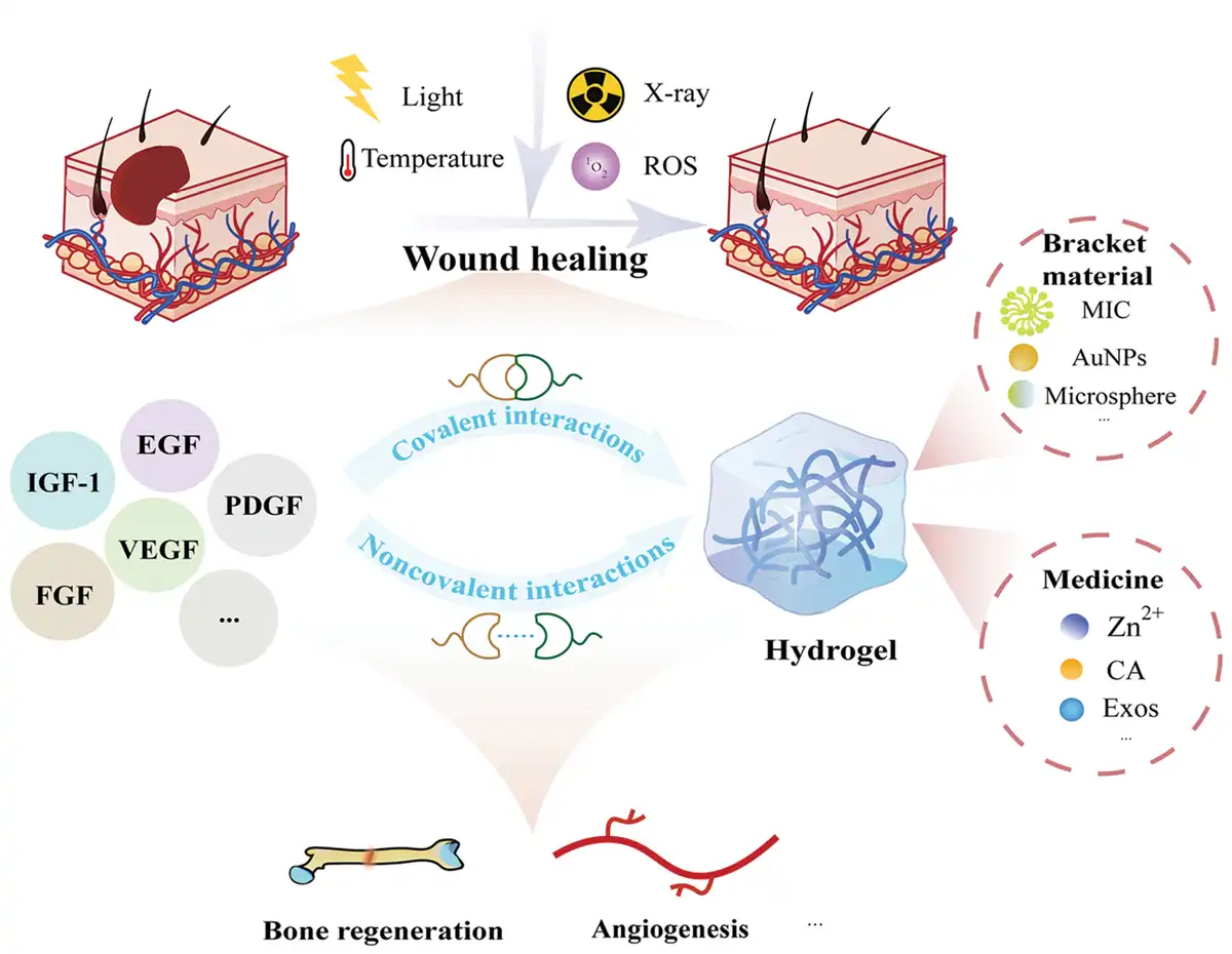
This review begins by examining key GFs employed in wound healing and their specific biological functions in tissue repair processes (Figure 1). Section 3 systematically details strategic methodologies for GF integration within hydrogels, encompassing both noncovalent encapsulation and covalent conjugation approaches, while further exploring the incorporation of stimuli-responsive elements to achieve spatiotemporally controlled release. Section 4 advances this foundation by introducing synergistic combination therapies that integrate GF-loaded systems with complementary therapeutic agents or bioactive scaffolds to potentiate wound regeneration. Section 5 subsequently evaluates the translational potential of these multifunctional hydrogels through their prospective applications in broader regenerative medicine contexts. Finally, we critically assess current technological limitations and emerging research trajectories, aiming to advance fundamental knowledge in wound healing biomaterials and establish robust frameworks for clinical translation.
Figure 1.
Scheme illustrating the use of hydrogel-based growth factor delivery systems for wound healing.
2. Growth factors for wound healing: classification and biological functions
The therapeutic application of GFs constitutes a major translational strategy in contemporary wound management. Key GFs that promote wound healing encompass vascular endothelial growth factor (VEGF), platelet-derived growth factor (PDGF), fibroblast growth factor (FGF), epidermal growth factor (EGF), and insulin-like growth factor-1 (IGF-1)[16]. This section systematically examines the mechanistic roles of these biomolecules in the wound healing cascade, as detailed in Figure 2.
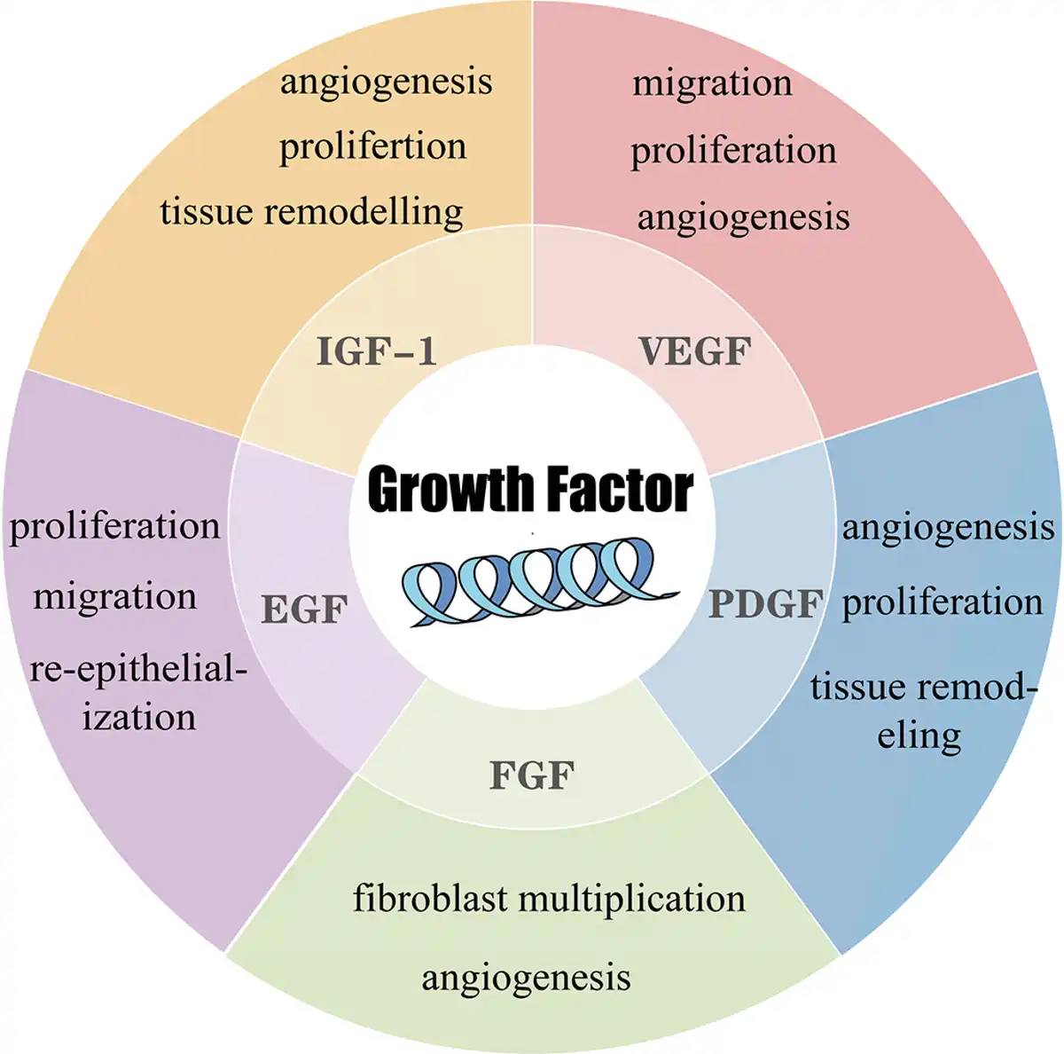
Figure 2.
Growth factors and their biological functions in wound healing. EGF, epidermal growth factor; FGF, fibroblast growth factor; IGF-1, insulin-like growth factor-1; PDGF, platelet-derived growth factor; VEGF, vascular endothelial growth factor.
2.1 Vascular endothelial growth factor
The VEGF family comprises 5 members: VEGF-A, VEGF-B, VEGF-C, VEGF-D, and placental growth factor[17]. VEGF-A binds to both VEGF-receptors (VEGFR)-1 and VEGFR-2 [18]. Given the endothelial-restricted expression of VEGFR-2, VEGF-A selectively activates endothelial cells to drive proliferative, migratory, angiogenic, and permeability-enhancing responses[17,19]. VEGF-C preferentially activates VEGFR-3 to stimulate lymphangiogenesis, whereas its binding to VEGFR-2 only marginally contributes to vascular permeability and angiogenesis[17,19]. By stimulating collagen (Col) production and accelerating re-epithelialization, VEGF serves as a multifunctional regulator in wound healing[9].
2.2 Platelet-derived growth factor
PDGF family consists of 5 isoforms formed by combinations of A- and B-polypeptide chains: PDGF-AA, PDGF-AB, PDGF-BB, PDGF-CC, and PDGF-DD[20]. PDGF-BB (becaplermin) was the first therapeutic GF agent approved by the US Food and Drug Administration for clinical treatment of chronic wounds[18]. PDGF is ubiquitously active throughout wound healing. Released from injured platelets, PDGF induces cellular proliferation, mediates chemotaxis of healing-associated cells (fibroblasts, macrophages, neutrophils, and monocytes), and ultimately drives extracellular matrix (ECM) regeneration[9,19]. Upon binding to its receptor, PDGF transmits mitogenic signals through 4 distinct pathways: (1) The tyrosine kinase cascade; (2) transcriptional activation of nuclear proto-oncogenes (c-Myc, c-Fos, and c-Jun); (3) receptor autophosphorylation and tyrosine phosphorylation of cytoplasmic substrates; and (4) intracellular Ca2+ elevation and Na+/H+ exchange activation[21]. Furthermore, PDGF contributes to tissue remodeling by upregulating matrix metalloproteinases (MMPs)[22].
2.3 Fibroblast growth factor
FGF family comprises 23 structurally related polypeptides, among which FGF-2, FGF-7, and FGF-10 serve as 3 crucial mediators in wound healing processes[19]. FGF-2 participates in all phases of wound healing[23]. It activates fibroblasts, vascular endothelial cells (VECs), smooth muscle cells, osteoblasts, and chondrocytes, while also enhancing keratinocyte motility[19,24]. Furthermore, FGF-2 accelerates re-epithelialization, enhances the synthesis and deposition of ECM components, and stimulates collagenase production[23,24]. Keratinocyte growth factor (KGF) is the common designation for FGF-7[25]. KGF is a critical mediator during the re-epithelialization phase, exerting paracrine effects exclusively through the FGFR2IIIb receptor expressed on keratinocytes[9,19]. KGF stimulates epithelial cell migration, proliferation, and differentiation, while also promoting Col deposition[9]. FGF-10 binds to FGFR1IIIb, exerting a mitogenic effect on cells expressing this receptor. Both FGF-7 and FGF-10 enhance the transcription of reactive oxygen species (ROS) detoxification-related factors, thereby protecting epithelial cells from damage under stressful conditions[19].
2.4 Epidermal growth factor
EGF is a 6-kDa endogenous polypeptide originally purified by Cohen from the murine submandibular gland[10]. The mitogenic EGF family comprises transforming growth factor-α (TGF-α), heparin-binding EGF-like growth factor (HB-EGF), amphiregulin, epiregulin, neuregulin, and betacellulin[18]. Among EGF family members, EGF, TGF-α, and HB-EGF serve as the primary mediators in wound healing processes[19]. Binding of these ligands to the epidermal growth factor receptor (EGFR) stimulates proliferation and migration of wound-associated keratinocytes[10,19]. EGF enhances wound healing through epidermal regeneration, keratinocyte/fibroblast activation, and granulation tissue-mediated contraction[16,26,27]. Furthermore, EGF serves as a critical regulator of epithelial cell motility and significantly accelerates re-epithelialization rates in diabetic wounds[28]. TGF-α enhances keratinocyte migration and proliferation while inducing expression of proliferation-associated keratins K6 and K16[19]. It contributes to both the initial stimulation and subsequent maintenance of wound re-epithelialization during early healing phases. However, studies have demonstrated that TGF-α deficiency exerts minimal impact on overall wound healing outcomes[18]. HB-EGF promotes re-epithelialization through binding to both EGFR (human epidermal growth factor receptor 1 [HER1]) and HER4 receptors[19]. HB-EGF functions as a mitogen and chemoattractant for fibroblasts and keratinocytes, suggesting its participation in granulation tissue induction[18,29].
2.5 Insulin-like growth factor-1
IGF-1, also known as somatomedin-C, is a peptide hormone predominantly produced by the liver under the control of growth hormone (GH)[30,31]. IGF-1 is a master regulator of somatic growth[32]. It binds to the IGF-1 receptor (IGF1R) in target cells to modulate downstream signaling cascades[9]. IGF-1 promotes wound healing by activating epidermal stem cells to regenerate the epithelium, stimulating proliferation and migration of fibroblasts and keratinocytes, and inducing vascularized granulation tissue formation[16,33]. Studies have demonstrated that IGF-1 suppresses inflammation in VECs through the Ras/PI3K/IKK/NF-κB signaling pathway while simultaneously promoting angiogenesis, thereby accelerating wound healing[33]. Furthermore, IGF-1 synergizes with other GFs to significantly accelerate the wound healing process[18].
3. Hydrogel combined with growth factors for wound healing
Hydrogels exhibit substantial potential in wound healing applications due to their inherent biocompatibility, high moisture retention capacity, and porous architecture that facilitates bioactive substance encapsulation. Building upon the review of GF mechanisms in Section 2, this section focuses on advanced strategies for synergizing hydrogels with GFs to potentiate wound repair efficacy. We systematically analyze: (1) methodologies for incorporating GFs into hydrogel matrices and (2) integration of stimuli-responsive modules for spatiotemporal control of release kinetics.
3.1 Growth factor incorporation strategies in hydrogels
The conjugation strategy employed for GF incorporation within hydrogels fundamentally dictates both bioactivity preservation and release kinetics. Integration approaches are principally categorized as noncovalent or covalent binding, each governing distinct release mechanisms: (1) degradation/erosion-controlled release, where therapeutic payload liberation coincides with hydrogel matrix dissociation; and (2) Diffusion-controlled release, involving aqueous permeation through the polymer network to solubilize and transport encapsulated agents[34]. This section systematically examines key methodologies for GF loading within hydrogel systems.
3.1.1 Non-covalent loading of growth factors in hydrogels
Noncovalent binding strategies encompass 3 primary approaches: physical encapsulation, electrostatic interactions, and affinity-based delivery. Critically, these strategies preserve the native structure and bioactivity of GFs by avoiding chemical modification. Among these methods, physical encapsulation involves the direct incorporation of either free GFs or GF-loaded nanoparticles (NPs) into the hydrogel matrix during network formation[35]. However, direct loading of GFs into hydrogel matrices typically results in rapid burst release. To mitigate this issue, Zuniga et al.[36] incorporated GFs into slowly-degrading keratin sulfate oligosaccharide (KSO) hydrogels. By adjusting the KSO concentration to reduce hydrogel porosity, they successfully prolonged the release duration. Notably, a 30% KSO concentration demonstrated the slowest release rate, sustaining GF release for 7 days.
Microspheres, small spherical particles ranging from 1 to 1000 μm in diameter, represent an alternative encapsulation platform for active compounds, including proteins, peptides, and GFs[37]. Seeking further extension of release kinetics, Xie et al.[38] developed a polyacrylamide (PAM) hydrogel system embedded with dual‑functional calcium alginate (ALG) microspheres. These microspheres were loaded with VEGF and coated with a polydopamine (PDA) layer, onto which silver nanoparticles (AgNPs) were conjugated. During the early inflammatory phase, the AgNPs release silver ions to exert antibacterial and anti-inflammatory effects, thereby helping to control infection and promote the transition to the proliferative phase. Concurrently, the acidic wound microenvironment during this phase helps retain the VEGF within the microspheres. As healing progresses into the proliferative phase and the microenvironment becomes more neutral, VEGF release is triggered, subsequently promoting angiogenesis, nutrient supply, and Col deposition. This composite microsphere-hydrogel system not only achieves sustained drug release but also delivers a time-programmed therapeutic action, ultimately accelerating infected wound healing[38]. While the aforementioned strategies achieve sustained release, they lack precise control over release kinetics. Addressing this limitation, Qin et al.[39] developed a hydrogel system employing direct PDGF incorporation. By varying the ratio of 2 crosslinkers to precisely modulate the hydrogel degradation rate, they achieved controlled regulation of PDGF release kinetics, as illustrated in Figure 3A[39].
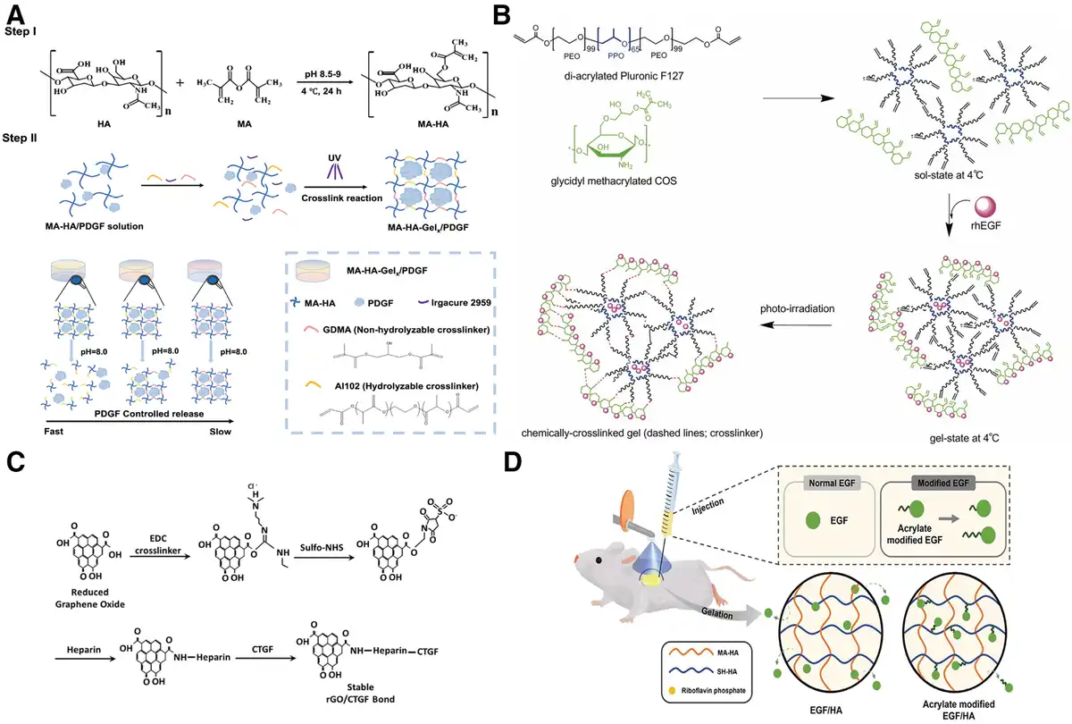
Figure 3.
Hydrogel-GF integration strategies. Noncovalent: (A) Physical entrapment[39]. Copyright 2025, Elsevier B.V. (B) Electrostatic interaction strategy[40]. Copyright 2010, Wiley Periodicals, Inc. (C) Affinity-based strategy (Heparin)[41]. Copyright 2024, Elsevier Ltd. (D) Covalent conjugation strategy[42]. Copyright 2023, American Chemical Society.
GFs can be electrostatically adsorbed onto hydrogel matrices via charge interactions between their surface properties and the hydrogel network. Leveraging this mechanism, Choi et al.[40] utilized the inherent negative charge of recombinant human epidermal growth factor (rhEGF, isoelectric point [pI] = 4.2) in neutral environments. They developed a pluronic/chitosan (CS) oligosaccharide (COS,molecular weight [MW] = 1–3 kDa, degree of deacetylation [DD] above 85%) hydrogel for rhEGF encapsulation by fabricating a physically crosslinked hydrogel combining glycidyl methacrylate-modified COS, diacrylated Pluronic, and rhEGF. Subsequently, Irgacure 2959 was introduced, and the mixture was exposed to long-wave ultraviolet (UV) light to establish chemical crosslinking (Figure 3B). This design leveraged ionic interactions between protonated amine groups on CS and the anionic rhEGF, achieving effective encapsulation. The resulting system facilitated targeted rhEGF release at the wound site, significantly accelerating wound healing[40].
Affinity-based loading surpasses physical encapsulation and electrostatic adsorption in preserving GF bioactivity while exhibiting superior binding affinity, consequently decelerating release kinetics. Heparin has emerged as a preeminent candidate for such delivery systems due to its anionic linear glycosaminoglycan structure and multifunctional pharmacological effects—including anticoagulation, antithrombotic, and anti-inflammatory activities that collectively potentiate wound healing[43–44–45]. Notably, the heparin released during the early treatment phase exerts its inherent anti-inflammatory effect, which helps modulate the wound microenvironment and thereby creates favorable conditions for subsequent repair. Crucially, conserved amino acid sequences in GFs serve as heparin-binding domains, with heparin conjugation preserving bioactivity and extending biological half-life[44,46,47]. Peng et al.[48] developed a hydrogel dressing composed of 4-arm acrylated polyethylene glycol (PEG) crosslinked with dithiothreitol, incorporating physically-bound heparin and basic FGF (bFGF). Experimental results demonstrated that this heparin-modified hydrogel enabled sustained bFGF release for at least 10 days. Exogenous bFGF not only directly promotes cell proliferation at the wound site but also upregulates the local expression of VEGF. Meanwhile, heparin maintains the patency of local microcirculation through its anticoagulant activity and synergizes with VEGF to amplify pro-angiogenic signaling, thereby significantly accelerating wound healing[48]. Rehman et al.[41] designed a gelatin (type A, from porcine skin) methacryloyl (GelMA) patch loaded with connective tissue growth factor (CTGF). Heparin was covalently conjugated to reduced graphene oxide (rGO) surfaces via amide bond formation, followed by CTGF addition to achieve heparin-mediated loading (Figure 3C). Notably, rGO itself has been reported to possess pro‑angiogenic potential and can enhance cell proliferation and migration, which may further synergize with the biological functions of CTGF and heparin. The resulting rGO/CTGF-incorporated hydrogel exhibited significantly reduced degradation rates compared with conventional hydrogels, maintaining 3D structural integrity for over 28 days[41]. Similarly, Zhao et al.[34] covalently crosslinked poly(guluronate) (PAG) from ALG (MW = 540 kDa, content of guluronic acid of about 70%). with aminated gelatin (AG, MW = 100 kDa type A, initial amino group content = 0.4903 mmol/g, amino group content after modification = 0.8438 mmol/g) to form PAG/AG hydrogels. Heparin was conjugated to gelatin through reductive amination, yielding heparin-grafted gelatin (HGC) for bFGF loading. The study compared bFGF release profiles from 3 systems: (1) hydrogels containing 2.0% free heparin (without gelatin), (2) hydrogels without HGC/heparin (direct bFGF loading), and (3) HGC-incorporated hydrogels with varied heparin ratios. Results indicated that the covalently conjugated HGC system achieved the slowest bFGF release, with release rates inversely proportional to heparin content[34]. Notably, covalently conjugated heparin-hydrogel matrices primarily release drugs through hydrogel degradation, whereas physically incorporated systems rely on diffusion-controlled release. Covalent conjugation thus enables substantially prolonged drug release compared with physical incorporation.
However, heparin exhibits limited binding specificity across different GFs, and excessive doses may induce bleeding complications[34,35]. Beyond heparin-based strategies, Adini et al.[49] developed a novel 12-amino acid peptide (PR1P) derived from an extracellular VEGF-binding domain. PR1P directly binds VEGF and potentiates its binding to endothelial cells. Yuan et al.[50] subsequently incorporated PR1P via physical adsorption into electrospun poly(L-lactide-co-glycolide)/gelatin (type B, from the porcine) (PG) fiber dressings to recruit endogenous VEGF and induce angiogenesis at wound sites[50]. However, this physical loading approach compromised PR1P functionality. To enhance VEGF recruitment efficiency, Zhang et al.[51] engineered a methacrylated hyaluronic acid (HA, MW = 90–100 kDa) (MeHA) hydrogel dressing with covalently conjugated PR1P. MeHA inherently supports angiogenesis and tissue regeneration, while its methacrylation enables stable hydrogel formation and controllable biodegradation. This system retained recruited VEGF and enabled controlled release through hydrogel degradation. Results confirmed that PR1P-conjugated hydrogels significantly promoted angiogenesis and accelerated wound healing[51]. Based on affinity-mediated GF loading, this approach prevents the loss of GF bioactivity while offering greater binding capacity and decelerated release kinetics compared with physical encapsulation or electrostatic interactions. This approach helps preserve GF bioactivity while generally offering greater binding capacity and decelerated release kinetics compared with physical encapsulation or electrostatic interactions.
3.1.2 Covalent loading of growth factors in hydrogels
Conventional hydrogel-based GF delivery frequently suffers from suboptimal pharmacokinetics, manifesting as initial burst release and inadequate therapeutic duration. Covalent immobilization within hydrogel matrices via chemical crosslinking presents a strategic solution to these limitations. Li et al.[52] pioneered an acrylate-functionalization approach for EGF, enabling covalent conjugation to HA networks. Methodologically, primary amine groups of EGF underwent derivatization with N-hydroxysuccinimide (NHS) esters to generate acrylate-EGF and acrylate-PEG-EGF conjugates; these were subsequently incorporated into thiol-modified HA (SH-HA, MW = 450000, degree of substitution [DS] = 75% ± 10%) matrices via thiol-ene click chemistry. Critically, in situ hydrogel formation occurred through thiol-ene crosslinking between MeHA (MW = 450000, DS = 75% ± 10%) and SH-HA (Figure 3D), establishing a molecularly engineered release platform. This design synchronizes EGF liberation with enzymatic hydrogel degradation, concurrently achieving sustained release kinetics and circumventing dose-dependent complications from uncontrolled EGF accumulation at wound sites[42].
3.2 Stimuli-responsive release systems
Conventional physical entrapment of GFs within hydrogels typically yields passive diffusion-controlled release kinetics, which often fails to satisfy the stringent therapeutic requirements for chronic wound management. In contrast, stimuli-responsive release systems enable precise spatiotemporal regulation of GF delivery, thereby orchestrating tissue regeneration through temporally coordinated molecular signaling. Critically, distinct GF incorporation methodologies dictate fundamentally different responsive release mechanisms. The subsequent discussion systematically examines stimulus-responsive release modules stratified by binding paradigms.
3.2.1 Stimuli-responsive release via noncovalent binding
Responsive release from physically encapsulated systems operates through structural disruption of hydrogel networks, inducing morphological relaxation to liberate GFs. Liu et al.[53] engineered a thermosensitive CS (MW = 50000–100000) hydrogel incorporating near-infrared (NIR)-absorbing PDANPs. NIR irradiation triggered localized photothermal conversion, prompting gel-to-sol phase transition and controlled human epidermal growth factor (hEGF) liberation (Figure 4A)[53]. Zhao et al.[54] exploited azobenzene photoisomerization within a supramolecular hydrogel, where UV-induced trans-to-cis transformation disrupted host-guest interactions, initiating network disassembly and EGF release. Precise dosage control was achieved through calibrated UV parameter modulation (Figure 4B)[54]. Xia et al.[55] demonstrated X-ray responsiveness via poly (β-amino ester) hydrogels containing disulfide bonds, MeHA (MW = 100000 kDa), and acrylamide radicals. X‑ray irradiation generated ROS, which in turn oxidized the disulfide bonds. This ROS-mediated oxidation induced network expansion and GF liberation (Figure 4C)[55]. The clinical application of X-ray-responsive systems is context-specific. It is primarily intended for managing radiation-induced skin injury in patients undergoing radiotherapy for cancer treatment. For the vast majority of chronic wounds not involving radiotherapy, the active application of X-rays is neither necessary nor aligned with clinical safety standards. In the context of chronic wound therapy, the clinical applicability of external energy-triggered approaches requires careful evaluation. UV radiation carries well-established risks of phototoxicity, immunosuppression, and potential carcinogenicity, along with limited tissue penetration[57]. While NIR light and low-energy visible light offer advantages in terms of penetration depth and biosafety, their energy delivery must still be strictly controlled to avoid thermal tissue damage. Endogenous microenvironment-responsive systems offer targeted alternatives: Jiang et al.[58] achieved mechanoresponsive PDGF release using N-phenylmethoxycarbonyl-L-tryptophan-modified hydrogels. Results demonstrated that under 0.1N tensile force, drug release from this mechanically responsive hydrogel increased approximately 2-fold compared with mechanically insensitive hydrogels[58]. Wang et al.[59] designed dual-responsive systems for diabetic wounds using phenylboronic acid-functionalized polylysine (PLL-PBA) and oxidized HA (OHA, MW = 1500000–2500000, degree of oxidation [DO] = 21.4%). The unoxidized O-diphenyl molecular structures within the hydrogel promote the aggregation and adhesion of red blood cells and platelets, thereby facilitating coagulation and hemostasis. Schiff base and boronic ester bonds underwent acid/ROS-triggered hydrolysis, enabling microenvironment-activated release. Notably, the cleavage of these dynamic covalent bonds consumes excess ROS and modulates local pH, while OHA, PLL-PBA, and the loaded drugs jointly exert synergistic antioxidant and anti-inflammatory effects. Subsequently, the released EGF acts to promote cellular proliferation and tissue regeneration, ultimately achieving sequential therapy that matches the natural healing progression[59].
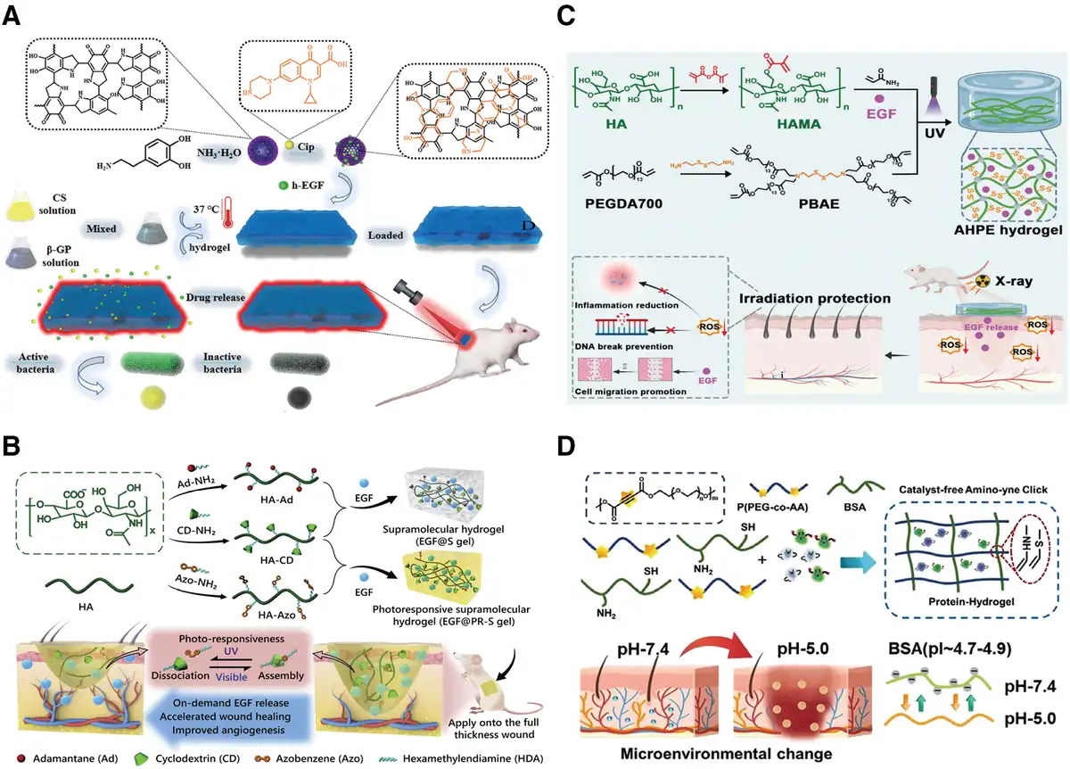
Figure 4.
Stimuli-responsive release mechanisms via noncovalent binding. (A) Thermoresponsive release[53]. Copyright 2021, Elsevier B.V. (B) Photo-responsive release[54]. Copyright 2020, Elsevier B.V. (C) X-ray-responsive release[55]. Copyright 2025, American Chemical Society. (D) pH-responsive release[56]. Copyright 2022, American Chemical Society.
Leveraging electrostatic interactions for controlled release, Zhang et al.[56] engineered a pH-responsive protein-based hydrogel system. The construct was fabricated through amino-alkyne bioconjugation between bovine serum albumin (BSA) and poly (ethylene glycol-co-acrylic acid) [P(PEG-co-AA)]. This design capitalizes on the divergent pI of BSA (pI 4.7–4.9) and bFGF (pI 9.1). Under physiological conditions (pH ≈ 7.4), protonation differences facilitate electrostatic complexation of cationic bFGF with anionic BSA chains. Critically, wound microenvironment acidification (pH ≈ 5.0) induced by hypoxia and bacterial proliferation approaches BSA’s pI, dramatically reducing its surface charge density. This electrostatic attenuation triggers bFGF liberation (Figure 4D). The system demonstrates intelligent spatiotemporal GF release responsive to pathological pH gradients, accelerating wound healing while establishing a novel therapeutic paradigm for chronic wound management[56]. Affinity-based loading technologies typically lack intrinsic stimuli-responsive mechanisms, thereby precluding precise spatiotemporal control over GF release kinetics without additional design elements.
3.2.2 Stimuli-responsive release via covalent conjugation
Covalent conjugation achieves GF liberation through stimuli-selective bond cleavage. Systems responsive to photonic, enzymatic, and oxidative stimuli enable spatiotemporally controlled release. Lin et al. engineered UV-cleavable EGF conjugates using heterobifunctional photocleavable (PC) linkers. The synthesis sequentially involved azido-PC-EGF derivation via NHS ester modification, dibenzocyclooctyne (DBCO)-HA preparation through thiol-maleimide conjugation, and EGF immobilization via strain-promoted alkyne-azide cycloaddition (SPAAC). Subsequent blue light-induced photopolymerization formed the final hydrogel (Figure 5A). Critically, UV-triggered linker cleavage enabled temporally controlled EGF release, maximizing therapeutic efficacy through precision dosing[60].
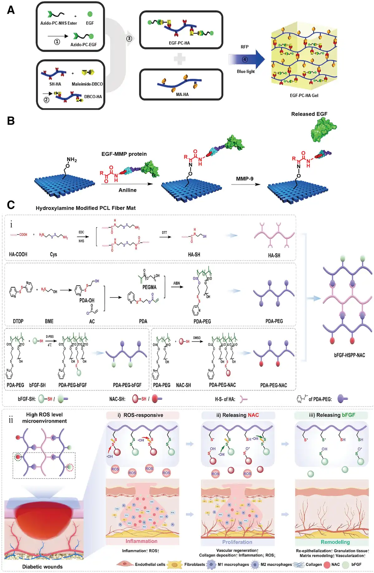
Chronic wounds exhibit characteristic pathological signatures, including MMP-9 upregulation and excessive ROS. Exploiting these biomarkers, researchers have engineered stimuli-responsive hydrogels capable of autonomous drug release without external triggers. Kim et al.[61] developed an MMP-9-responsive biological dressing through innovative protein engineering: First, they genetically incorporated an enzymatic cleavage site at the N-terminus of EGF, then performed pyridoxal 5’-phosphate-mediated transamination to introduce a ketone group. This functionalized EGF was conjugated via oxime bonds to microfibrous co-extruded poly(ε-caprolactone) (PCL) nonwoven felts, enabling protease-triggered release (Figure 5B). In the early phases of wound healing, MMP-9 is a key protease that is characteristically upregulated in the inflammatory microenvironment. This design ensures that EGF release is tightly coupled to the presence of this pathological signal, achieving spatiotemporally controlled delivery precisely when and where it is needed during the healing cascade[61]. Complementarily, Zheng et al.[62] designed a ROS-scavenging hydrogel comprising 3 functional components: PDA-PEG-bFGF, PDA-PEG-N-acetylcysteine (NAC), and HA-SH (a total thiol content of 72.1 µmol/g). Thiol-disulfide exchange reactions immobilized bFGF and NAC within the matrix. ROS exposure cleaves disulfide bonds into thiol groups, triggering the simultaneous release of both therapeutic agents (Figure 5C)[62]. Notably, the release of NAC occurs rapidly to suppress early inflammation and oxidative stress, thereby establishing a conducive microenvironment for the subsequent sustained action of bFGF, which in turn promotes cellular proliferation and ECM formation. This dual-action system concurrently alleviates oxidative stress while preventing ROS-mediated bFGF inactivation.
4. Combination therapy for wound healing
Although GF monotherapy may suffer from compromised bioactivity in proteolytic and inflammatory wound microenvironments, leading to diminished therapeutic outcomes, recent advances have engineered synergistic combination strategies. These integrate GFs with complementary therapeutics (e.g. antimicrobials and antioxidants) or nanostructured biomaterials (e.g., NPs and nanofibers) to orchestrate concurrent anti-inflammatory, angiogenesis-promoting, and (ECM) remodeling mechanisms for enhanced wound repair.
4.1 Synergistic therapy combining growth factors and pharmacological agents
Chronic wounds sustain pathophysiological microenvironments marked by ROS overload and recalcitrant bacterial colonization that impede healing cascades. Multifunctional hydrogels address these challenges through synergetic integration of GFs with complementary therapeutics, orchestrating coordinated tissue regeneration across distinct strategic paradigms. Xiong et al.[63] developed a multifunctional hydrogel co-loaded with MnO2 NPs and M2 macrophage-derived exosomes (M2 Exos) to synergize with FGF-2 for diabetic wound healing. The hydrogel was formed via Schiff base crosslinking between hydrazide-modified HA (HAh, MW = 200 kDa) and aldehyde-modified HA (HAa). MnO2 scavenged excess ROS/H2O2 to generate O2, alleviating oxidative stress and hypoxia, while M2 Exos promoted angiogenesis. This dual-action system enhanced cell proliferation, neovascularization, granulation tissue formation, and Col deposition, accelerating wound healing as shown in Figure 6A.
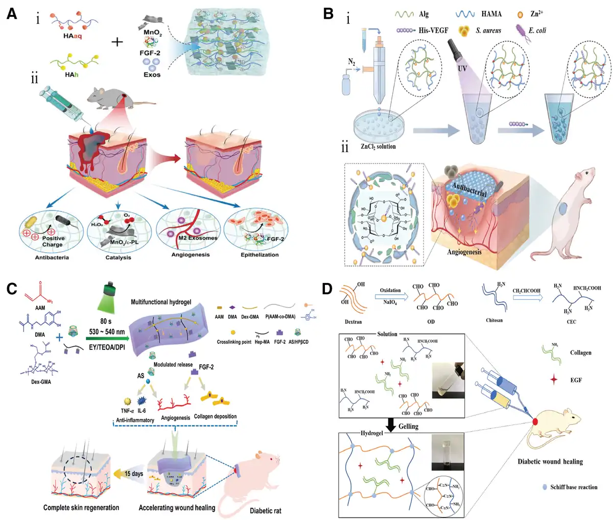
Beyond inorganic compounds, synthetic and natural organic compounds can be employed. Dopamine (DA) reduces ROS at wound sites while promoting M1-to-M2 macrophage polarization. Xie et al.[67] developed a hydrogel composed of oxidized dextran (OD, MW = 70000, DO = 83.16%), PLL, DA, and bFGF. This hydrogel exhibits ROS-scavenging, anti-inflammatory, epithelialization-promoting, and angiogenic properties, thereby accelerating wound healing; doxycycline (DOX), a semi-synthetic tetracycline, exhibits antibacterial, antioxidant, and anti-inflammatory properties. Gu et al.[68] developed a composite hydrogel co-loaded with DOX and FGF21, demonstrating combined antibacterial, antioxidant, anti-inflammatory, and pro-angiogenic functions to enhance wound recovery[68]; Metformin (Met), an antidiabetic drug, may modulate metabolic processes related to inflammation and oxidative damage while stimulating angiogenesis. Zhu’s team incorporated Met and FGF21 into a hydrogel composed of tsPBA and PVA for combined diabetic wound therapy[69].
Ju et al.[64] engineered a dual-functional hydrogel incorporating zinc ions (Zn2+) and VEGF[64]. Within this system, Zn2+ provided broad-spectrum antibacterial activity, while VEGF stimulated the proliferation and migration of VECs to enhance angiogenesis. This design synergistically combined sustained VEGF release via the microsphere carrier with Zn2+-mediated antibacterial effects, significantly accelerating wound healing (Figure 6B). Expanding beyond simple metal ion antibacterial strategies, Li et al.[52] exploited the photothermal properties of eutectic gallium-indium (EGaIn) liquid metal (LM). They developed an LM hybrid hydrogel co-loaded with VEGF, achieving wound healing acceleration through dual mechanisms: photothermally induced bacterial eradication and LM-enhanced angiogenesis. Similarly, leveraging LM technology, Wang et al.[70] integrated LM and EGF into a dialdehyde-carboxymethyl-cellulose (DCMC)-modified PAM hydrogel[70]. NIR irradiation triggered synergistic metal-photothermal therapy, providing potent antibacterial action while EGF concurrently promoted tissue regeneration and neovascularization. To enhance efficacy, Wang et al.[70] ultrasonically grafted copper onto LM, achieving 100% antimicrobial efficiency[71].
Natural products demonstrate synergistic potential with GFs for advanced wound healing. Kim et al.[72] co-encapsulated tannic acid (TA), a plant-derived polyphenol exhibiting potent antioxidant and anti-inflammatory properties, alongside PDGF within a thermosensitive poly (organophosphazene) hydrogel (TSP-TP). This system achieved combined therapeutic outcomes through ROS reduction, M2 macrophage polarization, and enhanced angiogenesis[72]. Similarly, Wang et al.[65] developed a dual-functional hydrogel via one-pot photopolymerization of dopamine methacrylamide, acrylamide, and glycidyl methacrylate-modified dextran (average MW = 70000, DS = 24.8%), co-incorporating asiaticoside (AS) and FGF-2. The resultant hydrogel exhibited concurrent antibacterial activity and promoted cellular proliferation/migration. In vivo validation confirmed accelerated wound healing via enhanced angiogenesis and Col deposition, as evidenced in Figure 6C[65].
ECM biomimetics establish barrier-proliferation codynamics: Hu et al. [66] developed an injectable hydrogel based on a Schiff base reaction between OD (MW = 100000 Da) and carboxylated CS (DD = 75–85%) (CEC). Crucially, Col, recognized as a critical component for both maintaining skin barrier function and promoting re-epithelialization, was incorporated into the hydrogel alongside EGF. Specifically, Col was immobilized within the hydrogel matrix through additional Schiff base reactions with CEC (Figure 6D). The synergistic action of Col and EGF significantly enhanced neotissue formation, improved Col deposition, and stimulated cell proliferation, collectively accelerating wound healing[66].
In combination therapies, drugs (such as antibacterial or anti-inflammatory components) act first to clean the wound and reduce inflammation, thereby creating a favorable local environment for repair. Subsequently, on the basis of this improved environment, GFs are released to directly promote key repair processes such as blood vessel growth and cell proliferation. We specifically distinguish the roles of different GFs: PDGF is primarily released during the early stages of healing. It recruits essential immune cells and repair cells to the wound site and works synergistically with early anti-inflammatory drugs to drive the transition of the wound from the inflammatory phase to the growth phase. In contrast, GFs such as VEGF and FGF focus on executing specific regenerative tasks, including building new blood vessels and promoting cell growth.
4.2 Synergistic therapy of growth factors and scaffold materials
In addition to synergistic therapy with drugs, GFs can also be combined with various functional scaffold materials for wound healing. Liu et al.[73] engineered a synergistic CS (MW = 100–150 kDa, DD = 85%)-HA hydrogel co-delivering FGF and gold nanoparticles (AuNPs) (Figure 7A). The system demonstrates dual functionality: AuNPs confer broad-spectrum antibacterial activity against MRSA, Staphylococcus aureus, Escherichia coli, and Pseudomonas aeruginosa, while FGF significantly enhances proliferation and migration of NIH/3T3-L1 and L929 fibroblasts. This combinatorial approach effectively promotes tissue regeneration while preventing bacterial infection, establishing promising therapeutic potential[73].
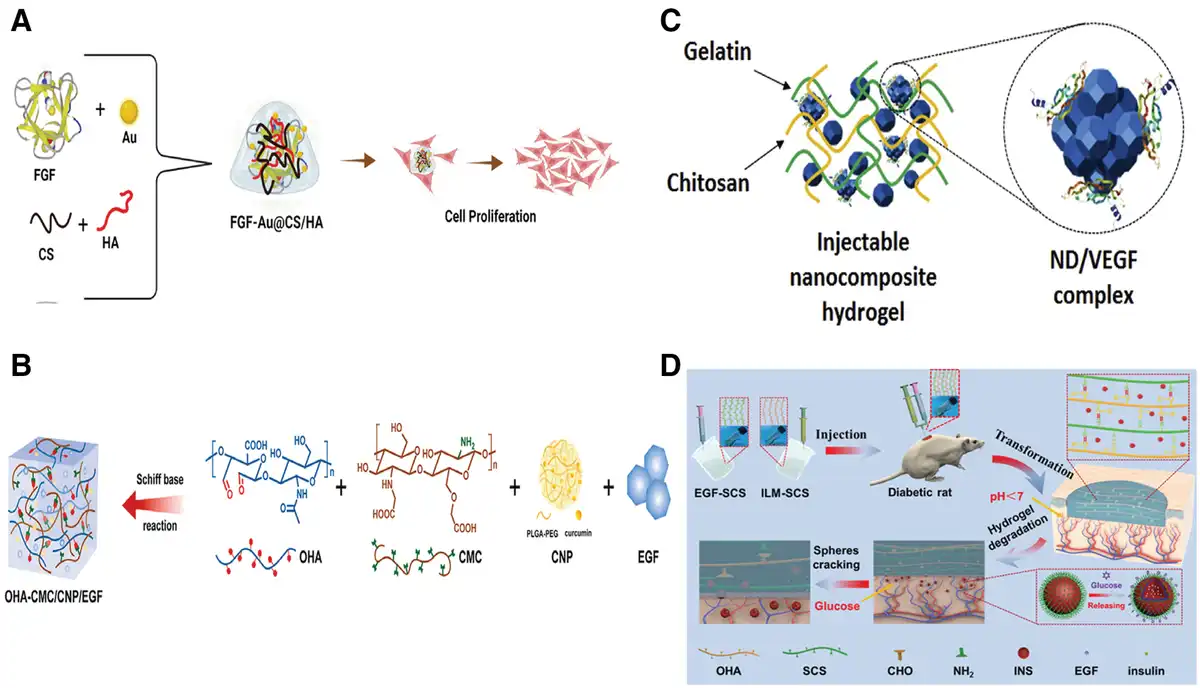
Hu et al.[74] developed a Schiff base-crosslinked OHA (MW = 100 kDa, DO = 56.0%)/carboxymethyl CS hydrogel co-encapsulating curcumin nanoparticles (CNPs) and EGF (Figure 7B). The design leverages EGF’s free amino groups for covalent conjugation, enabling sequential release kinetics: CNPs initially alleviate inflammation and oxidative stress, followed by EGF diffusion to promote cellular proliferation and migration. This temporal synchronization with diabetic healing phases provides an innovative regenerative strategy[74]. Pacelli et al.[75] innovated VEGF delivery via adsorption onto nanodiamonds (NDs) within a thermosensitive gelatin (type A, from porcine skin, bloom grade 300)/CS (MW = 10000 Da, low degree of acetylation [DA]) hydrogel dual-crosslinked with genipin/β-glycerophosphate (Figure 7C). NDs exhibit excellent biocompatibility while adsorbing and stabilizing VEGF, with the thermoresponsive matrix enabling precise implantation site localization. The system significantly prolongs VEGF release duration, achieving spatiotemporally controlled delivery for enhanced therapeutic efficacy[75]. Zhu et al.[76] synthesized glucose-responsive poly(ethylene glycol)-b-poly[3-acrylamidophenylboronic acid-co-styrene] micelles loaded with insulin (insulin-loaded micelles), incorporated with EGF into OHA (oxygenation efficiency = 52.5%)/succinyl CS (degree of acetylation ≥ 95%, DS = 15.4%) hydrogels (Figure 7D). PBA groups enable hyperglycemia-triggered micelle disassembly for insulin release, while EGF concurrently promotes epidermal proliferation. This dual-targeting mechanism demonstrates exceptional potential for accelerated chronic wound repair[76]. As with certain drug components that can improve the wound environment, many biomaterials themselves possess antibacterial, anti‑inflammatory, or cytoprotective functions. They can help control infection and alleviate inflammation before releasing GFs, thereby creating more favorable conditions for subsequent repair. The mechanism of action of NDs, however, differs. Studies have shown that NDs induce no significant cytotoxicity or adverse inflammatory responses in vitro and can form stable complexes with VEGF. This ensures sustained VEGF release while avoiding excessive inflammation triggered by the material itself, thereby enabling VEGF to focus on promoting the activation and proliferation of VECs and safeguarding the repair process from interference.
4.3 Synergistic therapy of hydrogel and scaffold materials
To address full-thickness skin defects, bilayer scaffolds mimicking epidermal and dermal matrices have been developed. Zandi et al.[77] engineered a bilayer scaffold featuring an epidermal layer of GelMA (type A, from porcine skin, bloom grade = 300)/laponite (LA) nanocomposite hydrogel with LA-immobilized EGF for prolonged release, and a dermal layer of glucose-crosslinked gelatin nanofibrous matrix. This design leverages glucose degradation for cellular energy while providing a nanofibrous architecture for cell proliferation and adhesion. The dermal layer absorbs exudate while the hydrogel maintains a moist microenvironment[77]. Song et al.[78] fabricated a scaffold comprising an electrospun PCL nanofibrous epidermal barrier loaded with amoxicillin for antibacterial protection, and a 3D-printed SA/Gel (type A, from porcine skin) dermal hydrogel encapsulating rhEGF to promote cellular processes and maintain moisture[78]. Expanding beyond nanofibers, Chen et al.[79] integrated inverse opal scaffolds with hydrogels for diabetic wounds. Their construct combines a N-acryloyl glycinamide/1-vinyl-1,2,4-triazole inverse opal scaffold providing structural integrity, antibacterial properties, and thermoresponsive color monitoring with a temperature-sensitive poly(N-isopropylacrylamide) hydrogel filler loaded with VEGF. Elevated temperatures trigger VEGF release to facilitate healing[79]. Collectively, these bilayer systems demonstrate superior efficacy over single-layer scaffolds in full-thickness wound repair.
5. Extended applications of growth factor-loaded hydrogels
Beyond wound healing, hydrogel-GF systems demonstrate therapeutic potential in diverse applications, including corneal regeneration, angiogenesis promotion, cervical cancer (CC) recurrence suppression, bone regeneration, and spinal cord injury (SCI) repair.
5.1 Corneal defect regeneration
Trauma-induced corneal defects can lead to corneal infection, ulceration, and scar formation, ultimately culminating in severe visual impairment[80]. Recent studies have demonstrated the therapeutic efficacy of GFs such as EGF and IGF-1 in promoting healing of corneal epithelial defects[81]. Hydrogel combined with GFs has consequently emerged as a promising strategy for managing corneal defects. Kang et al.[81] developed an in situ-forming bioorthogonal crosslinking hydrogel for treating corneal defects. In this system, EGF was conjugated to HA-PEG-DBCO using a PC linker, followed by the addition of azide-conjugated collagen (Col-N3). Subsequent hydrogel formation occurred in situ via a SPAAC reaction. As described in Section Stimuli-responsive release via covalent conjugation, EGF release could be triggered by UV irradiation[81]. The resulting hydrogel demonstrated the ability to fill corneal defects in situ without requiring exogenous initiators (e.g. light or heat) or catalysts, while exhibiting mechanical and biological properties comparable to those of the native corneal stroma[82]. The released EGF significantly promoted the proliferation and migration of corneal epithelial cells, thereby facilitating re-epithelialization (Figure 8A). These results collectively demonstrate that this in situ-formed, bioorthogonally crosslinked hydrogel incorporating releasable GFs represents a novel and promising therapeutic strategy for corneal defect repair[81].
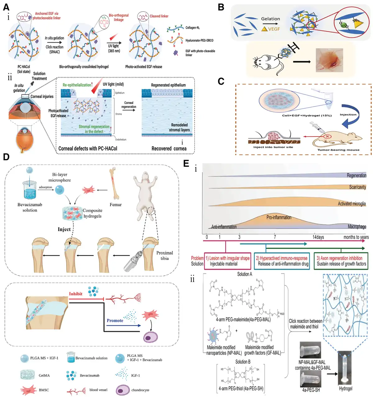
Figure 8.
Extended applications of hydrogel-GF systems. (A) Corneal defect regeneration[81]. Copyright 2024, The Authors. (B) Angiogenesis[83]. Copyright 2023, The Authors. (C) CC therapy[84]. Copyright 2024, Elsevier B.V. (D) Cartilage regeneration[85]. Copyright 2023, The Authors. (E) SCI repair[86]. Copyright 2021, Wiley-VCH GmbH.
5.2 Angiogenesis promotion
Vascular angiogenesis constitutes a complex, multistep biological process of pivotal importance in development, normal physiology, and disease pathology. However, successful reconstruction of functional vasculature in tissue engineering remains a formidable challenge[87]. As noted in Section Vascular endothelial growth factor, VEGF functions as a pro-angiogenic GF that plays a central role in both angiogenesis and vascular permeability regulation. Min et al.[83] incorporated VEGF into sulfated cellulose nanocrystal (CNC-S) hydrogels[83]. The positively charged VEGF molecules can bind to the negatively charged CNC-S primarily via electrostatic interactions. Critically, the sulfate groups on the CNC-S function as heparin-mimicking domains, which significantly enhance VEGF-binding affinity through specific molecular interactions (Figure 8B). Experimental results demonstrated that the CNC-S/VEGF hydrogel clearly promoted vascular generation. Consequently, this biomimetic system exhibits substantial potential for therapeutic vascularization applications. Furthermore, bFGF demonstrates potent angiogenic activity. Modaresifar et al.[88] encapsulated bFGF within GelMA (type A, from porcine skin, degree of methacrylation approaching 70%)/CS NPs composite hydrogels, enabling sustained release for efficient therapeutic delivery in wound healing and tissue regeneration applications[88]. Conjugation of pro-angiogenic GFs to hydrogel matrices preserves their bioactivity, representing an effective strategy for accelerating tissue regeneration.
5.3 Suppression of cervical cancer recurrence
In CC, overexpression of EGFR significantly contributes to tumor recurrence. To address this, Li et al.[84] developed a temperature-responsive hydrogel comprising a hydrophilic PEG block and a hydrophobic poly (lactic-co-glycolic acid) (PLGA) block (Figure 8C)[84]. Upon temperature elevation, intensified hydrophobic interactions within the PLGA segments drive the self-assembly of these amphiphilic block copolymers into a physically crosslinked hydrogel network. This in situ formed hydrogel inhibits recurrence through a dual mechanism: First, the encapsulation and subsequent temperature-triggered sustained release of EGF reduces local concentrations of free, bioactive EGF. Second, the inherent hydrophobic microenvironment facilitates the adsorption of excess EGF within the tumor niche, thereby diminishing its availability for binding to EGFR. Collectively, these actions suppress activation of the EGF-EGFR signaling pathway. Experimental results confirm that this hydrogel system significantly suppresses tumor growth, offering a viable clinical strategy for preventing CC recurrence.
5.4 Bone regeneration
Hydrogels functionalized with GFs offer significant potential for cartilage regeneration. Following injury, the intrinsically avascular and acellular nature of the growth plate severely limits its endogenous repair capacity, often resulting in the formation of bone bridges. These bony formations not only impede longitudinal skeletal growth but can also lead to significant deformities. To address this challenge, Qiang et al.[85] developed an innovative hydrogel system designed to prevent bone bridge formation and actively promote cartilage regeneration[85]. Specifically, this system integrates dual-compartment PLGA microspheres into a GelMA hydrogel encapsulating bone marrow-derived mesenchymal stem cells (BMSCs). Within these microspheres, the outer compartment is loaded with bevacizumab to inhibit aberrant vascularization and subsequent bone bridge formation, while the inner compartment contains IGF-1 to stimulate chondrogenic differentiation of BMSCs and facilitate cartilage regeneration (Figure 8D). Critically, this dual-compartment design enables the sequential release of bevacizumab followed by IGF-1, thereby providing a spatiotemporally controlled therapeutic approach. This strategy presents a novel dual-drug delivery platform for the effective treatment of growth plate cartilage injuries.
5.5 Spinal cord injury repair
Effective therapeutic approaches for SCI remain elusive, primarily due to its complex pathophysiological characteristics and limited endogenous repair capacity[89]. Addressing this challenge, Ye et al.[86] proposed a biomaterial design framework guided by 3 core principles: (1) injectability coupled with high biocompatibility and controlled degradability; (2) precise spatiotemporal release of anti-neuroinflammatory drugs; and (3) sustained long-term delivery of essential GFs during the chronic repair phase. Translating this rationale into practice, they engineered an in situ self-assembling hydrogel co-loaded with therapeutic agents. Methylprednisolone sodium succinate (MPSS) was encapsulated in NPs. The NPs were surface-functionalized with polymer acrylate-PEG-NHS and then reacted with 1-(2-aminoethyl)-1H-pyrrole-2,5-dione hydrochloride (NH2-MAL) via urethane condensation, yielding NP-MAL. Concurrently, GFs were maleimide-modified (GF-MAL). Hydrogel network formation occurred via thiol-maleimide click chemistry between NP-MAL, GF-MAL, and 4a-PEG-thiol. This design confers distinct therapeutic advantages: Encapsulating MPSS within NPs prolonged its release profile, effectively mitigating acute inflammatory responses and preventing postinjury cavity formation. Conversely, covalent conjugation of GFs via the maleimide linkers enabled their sustained, long-term release, crucial for enhancing neuron survival and tissue repair throughout the chronic phase (Figure 8E). Collectively, this multifunctional hydrogel platform provides a rationally designed and effective biomaterial strategy for acute SCI intervention[86].
6. Summary and outlook
Hydrogel-GF composites exhibit considerable translational potential in wound management. While noncovalent conjugation offers procedural simplicity under mild conditions, its weak binding affinity predisposes systems to burst release kinetics. Conversely, covalent immobilization minimizes initial burst release but may compromise GF bioactivity through chemical modification and requires more complex synthesis. Critically, both conjugation paradigms enable stimuli-responsive release control. Photolabile hydrogels achieve spatiotemporal precision via PC linker dissociation (e.g. o-nitrobenzyl ester cleavage for EGF liberation), whereas pathological microenvironment-responsive systems (ROS/MMP-9 activated) leverage endogenous triggers to overcome limitations associated with exogenous stimuli. To optimize the healing milieu, multifunctional hydrogels integrate GFs with complementary therapeutics (antimicrobials, immunomodulators) and structural scaffolds, synergistically addressing infection control, proliferation potentiation, and matrix remodeling. Beyond cutaneous repair, this technology demonstrates versatility in osteogenesis, spinal cord regeneration, and corneal defect restoration.
Although preclinical studies validate system efficacy in epithelial regeneration, inflammation suppression, and cellular differentiation, clinical translation confronts persistent challenges. Future advancements should focus on: (1) development of multiresponsive smart materials: integrating multiple responsive mechanisms (light, temperature, pH, enzymes, etc.) to achieve cascaded release of GFs, precisely matching the multi-stage needs of wound healing (inflammatory phase, proliferative phase, remodeling phase); (2) development of low-cost processes: employing 3D printing and microfluidic techniques to achieve personalized customization and batch production of hydrogels; (3) interdisciplinary collaborative research: utilizing artificial intelligence (AI) to predict GF release kinetics and customize GF formulation strategies and release profiles based on patient-specific parameters (e.g. wound type, metabolic status) to optimize material design and treatment protocols.
In summary, the strategic convergence of hydrogels and GFs, coupled with synergistic combinations and intelligent release mechanisms, will propel wound care from empirical management toward precision medicine. This evolution points toward a potential prospect of achieving comprehensive regeneration for complex wounds through single-intervention strategies.
Acknowledgements
This work was financially supported by the Key Research Development Program of Zhejiang Province (Grant No. 2024C03012), the National Natural Science Foundation of China (Grant Nos. 22408078, 82401057, and 32101170), and Zhejiang Province Postdoctoral Excellence Funding Program-Special Support (Grant No. ZJ2024004).
Conflicts of interests
The authors declare that they have no conflicts of interest.
Author contributions
Yawen Xue drafted the initial manuscript. Kan Zhan reviewed and edited the manuscript. The investigation was conducted by Junping Zhou and Yanan Xue. Liqun Jin, Renchao Zheng, and Yuguo Zheng supervised the manuscript and project. All authors reviewed the manuscript.
References
- [1] Tchemtchoua VT, Atanasova G, Aqil A, et al. Development of a chitosan nanofibrillar scaffold for skin repair and regeneration. Biomacromolecules. 2011;12(9):3194–3204.
- [2] Eke G, Mangir N, Hasirci N, et al. Development of a UV crosslinked biodegradable hydrogel containing adipose derived stem cells to promote vascularization for skin wounds and tissue engineering. Biomaterials. 2017;129:188–198.
- [3] Han G, Ceilley R. Chronic wound healing: a review of current management and treatments. Adv Ther. 2017;34(3):599–610.
- [4] Qi LF, Zhang CL, Wang B, et al. Progress in hydrogels for skin wound repair. Macromol Biosci. 2022;22(7):e2100475.
- [5] Ongarora BG. Recent technological advances in the management of chronic wounds: a literature review. Health Sci Rep. 2022;5(3):e641.
- [6] Sen CK. Human wound and its burden: Updated 2020 compendium of estimates. Adv Wound Care (New Rochelle). 2021;10(5):281–292.
- [7] Xu ZJ, Han SY, Gu ZP, et al. Advances and impact of antioxidant hydrogel in chronic wound healing. Adv Healthcare Mater. 2020;9(5):e1901502.
- [8] Shan BH, Wu FG. Hydrogel-based growth factor delivery platforms: strategies and recent advances. Adv Mater. 2024;36(5):e2210707.
- [9] Zubair M, Ahmad J. Role of growth factors and cytokines in diabetic foot ulcer healing: a detailed review. Rev Endocr Metab Disord. 2019;20(2):207–217.
- [10] Shakhakarmi K, Seo JE, Lamichhane S, et al. EGF, a veteran of wound healing: highlights on its mode of action, clinical applications with focus on wound treatment, and recent drug delivery strategies. Arch Pharm Res. 2023;46(4):299–322.
- [11] Xu YM, Hu QY, Wei ZY, et al. Advanced polymer hydrogels that promote diabetic ulcer healing: mechanisms, classifications, and medical applications. Biomater Res. 2023;27(1):36.
- [12] Dong RN, Guo BL. Smart wound dressings for wound healing. Nano Today. 2021;41:101290.
- [13] Zhang T, Liu F, Tian W. Advance of new dressings for promoting skin wound healing. Sheng Wu Yi Xue Gong Cheng Xue Za Zhi. 2019;36(6):1055–1059.
- [14] Niculescu AG, Grumezescu AM. An up-to-date review of biomaterials application in wound management. Polymers. 2022;14(3):421.
- [15] Zheng HY, Zhang XF, Wang WJ, et al. Oxidized hyaluronic acid/ε-polylysine/quaternized chitosan hydrogel with shear thinning injectability and self-healing properties for full-time and multipurpose wound healing. Int J Biol Macromol. 2025;293:139347.
- [16] Yadav JP, Singh AK, Grishina M, et al. Insights into the mechanisms of diabetic wounds: pathophysiology, molecular targets, and treatment strategies through conventional and alternative therapies. Inflammopharmacology. 2024;32(1):149–228.
- [17] Saaristo A, Tammela T, Farkkila A, et al. Vascular endothelial growth factor-c accelerates diabetic wound healing. Am J Pathol. 2006;169(3):1080–1087.
- [18] Werner S, Grose R. Regulation of wound healing by growth factors and cytokines. Physiol Rev. 2003;83(3):835–870.
- [19] Barrientos S, Stojadinovic O, Golinko MS, et al. Growth factors and cytokines in wound healing. Wound Repair Regen. 2008;16(5):585–601.
- [20] Zhang M, Yu W, Niibe K, et al. The effects of platelet-derived growth factor-BB on bone marrow stromal cell-mediated vascularized bone regeneration. Stem Cells Int. 2018;2018:3272098.
- [21] Zhang ZM. Analysis of perioperative myocardial ischemia and cardiac events in patients with myocardial infarction who underwent noncardiac surgery. Int Med Health Guidance News. 2002;8(7):46–48.
- [22] Sun XJ, Gao X, Zhou LY, et al. PDGF-BB-induced MT1-MMP expression regulates proliferation and invasion of mesenchymal stem cells in 3-dimensional collagenMEK/ERK1/2 and PI3K/AKT signaling. Cell Signal. 2013;25(5):1279–1287.
- [23] Syromiatnikova VY, Kvon AI, Starostina IG, et al. Strategies to enhance the efficacy of FGF2-based therapies for skin wound healing. Arch Dermatol Res. 2024;316(7):405.
- [24] Koike Y, Yozaki M, Utani A, et al. Fibroblast growth factor 2 accelerates the epithelial-mesenchymal transition in keratinocytes during wound healing process. Sci Rep. 2020;10(1):18545.
- [25] Wang R, An K, Xie J, et al. [Role of fibroblast growth factor 7 in craniomaxillofacial development]. Sichuan Da Xue Xue Bao Yi Xue Ban. 2024;55(2):469–474.
- [26] Hardwicke J, Schmaljohann D, Boyce D, et al. Epidermal growth factor therapy and wound healing—past, present and future perspectives. Surgeon. 2008;6(3):172–177.
- [27] Dogan S, Demirer S, Kepenekci I, et al. Epidermal growth factor-containing wound closure enhances wound healing in non-diabetic and diabetic rats. Int Wound J. 2009;6(2):107–115.
- [28] Bodnar RJ. Epidermal growth factor and epidermal growth factor receptor: The yin and yang in the treatment of cutaneous wounds and cancer. Adv Wound Care (New Rochelle). 2013;2(1):24–29.
- [29] Vaidyanathan L. Growth factors in wound healing – a review. Biomed Pharmacol J. 2021;14(3):1469–1480.
- [30] Yoshida T, Delafontaine P. Mechanisms of IGF-1-mediated regulation of skeletal muscle hypertrophy and atrophy. Cells. 2020;9(9):1970.
- [31] Böhm M, Stegemann A, Paus R, et al. Endocrine controls of skin aging. Endocr Rev. 2025;46(3):349–375.
- [32] Clemmons DR. Role of IGF binding proteins in regulating metabolism. Trends Endocrinol Metab. 2016;27(6):375–391.
- [33] Zhang X, Hu F, Li J, et al. IGF-1 inhibits inflammation and accelerates angiogenesis via RAS/PI3K/IKK/NF-kappaB signaling pathways to promote wound healing. Eur J Pharm Sci. 2024;200:106847.
- [34] Zhao YL, Lin ZZ, Liu WQ, et al. Controlled release of growth factor from heparin embedded poly(aldehyde guluronate) hydrogels and its effect on vascularization. Gels. 2023;9(7):589.
- [35] Chen T, Jiang Y, Huang JP, et al. Essential elements for spatiotemporal delivery of growth factors within bio-scaffolds: a comprehensive strategy for enhanced tissue regeneration. J Control Release. 2024;368:97–114.
- [36] Zuniga K, Isaac A, Christy S, et al. Characterization of a human platelet lysate-loaded keratin hydrogel for wound healing applications in vitro. Int J Mol Sci. 2022;23(8):4100.
- [37] Alghamdi A, Alissa M, Alshehri MA. Hydrogel based on adipose tissue-derived extracellular matrix encapsulated with microspheres containing epidermal growth factor (EGF) and vascular endothelial growth factor (VEGF) accelerate diabetic wound healing in a rat model. Bratisl Med J. 2025;126(6):2811–2813.
- [38] Xie XF, Lei H, Fan DD. Antibacterial hydrogel with pH-responsive microcarriers of slow-release VEGF for bacterial infected wounds repair. J Mater Sci Technol. 2023;144:198–212.
- [39] Qin XY, Yuan HX, Zhang M, et al. Controlled platelet-derived growth factor delivery by hyaluronic acid hydrogels with tunable crosslinking for accelerated chronic wound healing. Int J Biol Macromol. 2025;304(Pt 1):140710.
- [40] Choi JS, Yoo HS. Pluronic/chitosan hydrogels containing epidermal growth factor with wound-adhesive and photo-crosslinkable properties. J Biomed Mater Res A. 2010;95A(2):564–573.
- [41] Rehman SRU, Ullah A, Augustine R, et al. Enhanced wound healing by nanoengineered hydrogel patch loaded with connective tissue growth factor. Mater Today Commun. 2024;40:109825.
- [42] Lee YR, Lim S, Kim JA, et al. Development of thiol-ene reaction-based ha hydrogel with sustained release of EGF for enhanced skin wound healing. Biomacromolecules. 2023;24(11):5342–5352.
- [43] Gupta S, Puttaiahgowda YM, Deiglmayr L. Recent advances in the design and immobilization of heparin for biomedical application: a review. Int J Biol Macromol. 2024;264:130743.
- [44] Tan Q, Tang H, Hu JG, et al. Controlled release of chitosan/heparin nanoparticle-delivered VEGF enhances regeneration of decellularized tissue-engineered scaffolds. Int J Nanomedicine. 2011;6:929–942.
- [45] Qiu M, Huang SJ, Luo CH, et al. Pharmacological and clinical application of heparin progress: an essential drug for modern medicine. Biomed Pharmacother. 2021;139:111561.
- [46] Chung HJ, Kim HK, Yoon JJ, et al. Heparin immobilized porous PLGA microspheres for angiogenic growth factor delivery. Pharm Res. 2006;23(8):1835–1841.
- [47] Du P, Diao L, Lu YC, et al. Heparin-based sericin hydrogel-encapsulated basic fibroblast growth factor for in vitro and in vivo skin repair. Heliyon. 2023;9(3):e13554.
- [48] Peng J, Zhao H, Tu CZ, et al. Hydrogel dressing loaded with heparin and basic fibroblast growth factor for accelerating wound healing in rat. Mater Sci Eng C Mater Biol Appl. 2020;116:111169.
- [49] Adini A, Adini I, Chi ZL, et al. A novel strategy to enhance angiogenesis in vivo using the small VEGF-binding peptide PR1P. Angiogenesis. 2017;20(3):399–408.
- [50] Yuan ZC, Shafiq M, Zheng H, et al. Multi-functional fibrous dressings for infectious injury treatment with anti-adhesion wound healing. Mater Design. 2023;235:112459.
- [51] Zhang W, Qian SY, Chen J, et al. Photo-crosslinked pro-angiogenic hydrogel dressing for wound healing. Int J Mol Sci. 2024;25(18):9948.
- [52] Li JB, Wang Y, Fan L, et al. Liquid metal hybrid antibacterial hydrogel scaffolds from 3D printing for wound healing. Chem Eng J. 2024;496:153805.
- [53] Liu SP, Liu ZC, Wu MY, et al. Nir as a “trigger switch” for rapid phase change, on-demand release, and photothermal synergistic antibacterial treatment with chitosan-based temperature-sensitive hydrogel. Int J Biol Macromol. 2021;191:344–358.
- [54] Zhao W, Li Y, Zhang X, et al. Photo-responsive supramolecular hyaluronic acid hydrogels for accelerated wound healing. J Control Release. 2020;323:24–35.
- [55] Xia Y, Gui H, Li X, et al. X-ray responsive antioxidant drug-free hydrogel for treatment of radiation skin injury. ACS Appl Mater Interfaces. 2025;17(4):5671–5683.
- [56] Zhang R, Tian YC, Pang L, et al. Wound microenvironment-responsive protein hydrogel drug-loaded system with accelerating healing and antibacterial property. ACS Appl Mater Interfaces. 2022;14(8):10187–10199.
- [57] Gromkowska-Kepka KJ, Puscion-Jakubik A, Markiewicz-Zukowska R, et al. The impact of ultraviolet radiation on skin photoaging - review of in vitro studies. J Cosmet Dermatol. 2021;20(11):3427–3431.
- [58] Jiang HY, Lu X, Bu TS, et al. Mechanics mediated semi-convertible hydrogel enabled sustained drug release. Adv Healthc Mater. 2025;14(18):e2500845.
- [59] Wang SQ, Liu YH, Wang XS, et al. Modulating macrophage phenotype for accelerated wound healing with chlorogenic acid-loaded nanocomposite hydrogel. J Control Release. 2024;369:420–443.
- [60] Lim S, Kim JA, Chun YH, et al. Hyaluronic acid hydrogel for controlled release of heterobifunctional photocleavable linker-modified epidermal growth factor in wound healing. Int J Biol Macromol. 2023;253:126603.
- [61] Kim SE, Lee PW, Pokorski JK. Biologically triggered delivery of EGF from polymer fiber patches. ACS Macro Lett. 2017;6(6):593–597.
- [62] Zheng MH, Song WX, Huang PP, et al. Drug conjugates crosslinked bioresponsive hydrogel for combination therapy of diabetic wound. J Control Release. 2024;376:701–716.
- [63] Xiong Y, Chen L, Liu P, et al. All-in-one: Multifunctional hydrogel accelerates oxidative diabetic wound healing through timed-release of exosome and fibroblast growth factor(vol 18, 2104229, 2022). Small. 2023;19(21):e2104229.
- [64] Ju YK, Zeng HX, Ye XZ, et al. Zn2+ incorporated composite polysaccharide microspheres for sustained growth factor release and wound healing. Mater Today Bio. 2023;22:100739.
- [65] Wang Z, Zhao FZ, Deng JX, et al. Photopolymerized multifunctional hydrogel loaded with asiaticoside and growth factor for accelerating diabetic wound healing. Acs Appl Polym Mater. 2024;6(7):4267–4281.
- [66] Hu P, Lei QQ, Duan SX, et al. In-situ formable dextran/chitosan-based hydrogels functionalized with collagen and egf for diabetic wounds healing. Biomater Adv. 2022;136:212773.
- [67] Xie HL, Tian SB, Cui C, et al. A glycopeptide-based pH-responsive hydrogel promotes diabetic wound healing via antimicrobial and remodeling microenvironment. Colloids Surf B Biointerfaces. 2025;251:114614.
- [68] Gu YN, Liang Z, Wang YP, et al. Engineered adscs and doxycycline encapsulated antibacterial, immunomodulatory and proangiogenic hydrogels for infected burn wound healing. Chem Eng J. 2025;503:158478.
- [69] Zhu H, Xu J, Zhao M, et al. Adhesive, injectable, and ROS-responsive hybrid polyvinyl alcohol (PVA) hydrogel co-delivers metformin and fibroblast growth factor 21 (FGF21) for enhanced diabetic wound repair. Front Bioeng Biotechnol. 2022;10:968078.
- [70] Wang B, Fan LL, Cui ZL, et al. Liquid-metal-loaded hydrogel with antibacterial effect and sustained release of growth factor for wound healing. Adv Therap. 2024;7(6):2400056.
- [71] Wang B, Shan XH, Gao JY, et al. 3D-printed hydrogel patches embedded with Cu-modified liquid metal nanoparticles for accelerated wound healing. Adv Healthc Mater. 2025;14(15):e2404986.
- [72] Kim J, Bong KW, Cho JK, et al. Thermo-responsive hydrogel sustained co-delivery of TA and PDGF to modulate the diabetic microenvironment and accelerate diabetic wound healing. J Mater Chem B. 2025;13(24):7090–7105.
- [73] Liu X, Peng SW, Pei YJ, et al. Facile fabrication of chitosan/hyaluronic acid hydrogel-based wound closure material co-loaded with gold nanoparticles and fibroblast growth factor to improve anti-microbial and healing efficiency in diabetic wound healing and nursing care. Regen Ther. 2024;26:1018–1029.
- [74] Hu B, Gao MZ, Boakye-Yiadom KO, et al. An intrinsically bioactive hydrogel with on-demand drug release behaviors for diabetic wound healing. Bioact Mater. 2021;6(12):4592–4606.
- [75] Pacelli S, Acosta F, Chakravarti AR, et al. Nanodiamond-based injectable hydrogel for sustained growth factor release: preparation, characterization and analysis. Acta Biomater. 2017;58:479–491.
- [76] Zhu JY, Jiang GH, Hong WJ, et al. Rapid gelation of oxidized hyaluronic acid and succinyl chitosan for integration with insulin-loaded micelles and epidermal growth factor on diabetic wound healing. Mater Sci Eng C Mater Biol Appl. 2020;117:111273.
- [77] Zandi N, Dolatyar B, Lotfi R, et al. Biomimetic nanoengineered scaffold for enhanced full-thickness cutaneous wound healing. Acta Biomater. 2021;124:191–204.
- [78] Song YT, Hu QX, Liu SH, et al. Electrospinning/3D printing drug-loaded antibacterial polycaprolactone nanofiber/sodium alginate-gelatin hydrogel bilayer scaffold for skin wound repair. Int J Biol Macromol. 2024;275:129705.
- [79] Chen CW, Wang Y, Zhang H, et al. Responsive and self-healing structural color supramolecular hydrogel patch for diabetic wound treatment. Bioact Mater. 2022;15:194–202.
- [80] Gong JJ, Ding G, Hao ZK, et al. Elucidating the mechanism of corneal epithelial cell repair: Unraveling the impact of growth factors. Front Med (Lausanne). 2024;11:1384500.
- [81] Kang NW, Seo YA, Jackson KJ, et al. Photoactivated growth factor release from bio-orthogonally crosslinked hydrogels for the regeneration of corneal defects. Bioact Mater. 2024;40:417–429.
- [82] Chen F, Le P, Fernandes-Cunha GM, et al. Bio-orthogonally crosslinked hyaluronate-collagen hydrogel for suture-free corneal defect repair. Biomaterials. 2020;255:120176.
- [83] Min K, Tae G. Cellular infiltration in an injectable sulfated cellulose nanocrystal hydrogel and efficient angiogenesis by VEGF loading. Biomater Res. 2023;27(1):28.
- [84] Piao L, Xiang P, Zhou YY, et al. Thermo-sensitive PLGA-PEG-PLGA hydrogel for sustained release of EGF to inhibit cervical cancer recurrence. Colloids Surf B Biointerfaces. 2024;236:113795.
- [85] Qiang L, Fan MJ, Wang YW, et al. Injectable hydrogel loaded with bilayer microspheres to inhibit angiogenesis and promote cartilage regeneration for repairing growth plate injury. Front Bioeng Biotechnol. 2023;11:1181580.
- [86] Ye JJ, Jin S, Cai WX, et al. Rationally designed, self-assembling, multifunctional hydrogel depot repairs severe spinal cord injury. Adv Healthc Mater. 2021;10(13):e2100242.
- [87] Gao ZH, Yu YM, Dai K, et al. Engineering neutrophil immunomodulatory hydrogels promoted angiogenesis. ACS Appl Mater Interfaces. 2022;14(35):39746–39758.
- [88] Modaresifar K, Hadjizadeh A, Niknejad H. Design and fabrication of GelMA/chitosan nanoparticles composite hydrogel for angiogenic growth factor delivery. Artif Cells Nanomed Biotechnol. 2018;46(8):1799–1808.
- [89] Wang WZ, Sun MJ, Zhang ZD, et al. Application of microenvironment biomimetic materials in spinal cord injury and future prospects. Med Mat. 2025;2(3):e00020.
This article is available under Open Access.
© 2026 The Authors